Hand / Foot
Contact Person
- OÄ Dr. med. K. Schenk
- OA Herr J. Lietz
Focus Magazine Doctor's List 2016
Special Consultation Hours
Surgery and treatment methods of the foot
- Upper ankle total joint arthroplasty (TEP)
- Upper ankle joint TEP replacement
- Medial foot edge disentanglement
- Midfoot and hindfoot realignment osteotomies (e.g. wedge osteotomy)
- Hallux valgus operations
- Hallux rigidus operations / big toe metatarsophalangeal joint prostheses
- Osteotomies with joint preservation or resection Arthrodesis
- Soft tissue interventions
- Toe corrections for hammer and claw toes
- Surgery according to Hohmann
- Weil's osteotomies
Surgery and Treatment Methods Wrist and Forearm
- Osteosyntheses of the radius and ulna
- Lengthening and shortening osteotomies
- Arthodeses
- Synovectomies
- Epiphysiodesis
- Tenolyses
- Tenosynovectomies
- Radiosynoriontheses (RSD)
Patient Information on Selected Surgical and Treatment Methods
Cause of Ssteoarthritis
Pain at night and at rest is also possible here, and occasionally swelling may also occur in the area of the joint.
Osteoarthritis of the upper ankle joint can have various causes, such as post-traumatic changes (e.g. after fractures) or in the context of inflammatory joint diseases such as rheumatoid arthritis. In some cases, however, no actual cause for the joint wear will be found.
Treatment options Arthrodesis
Similar to the healing of a bone fracture, the joint then grows together. By removing bone wedges, it is also possible here to correct malpositions in the upper ankle joint accordingly.
With a fusion of the upper ankle joint, pain can be greatly reduced in most patients. Part of the lost mobility can be compensated for by the adjacent joints in the midfoot, so that there are often no significant restrictions on normal walking.
Possible Complications
However, there are also some known disadvantages of stiffening. For example, a long period of immobilization of the joint (up to 100 days) is generally required after the operation until the joint is finally fused. In up to 20% of cases, bone healing may also be delayed or may not occur at all. Then we must contend with, a so-called pseudarthrosis, which then entails one or more subsequent operations.
Furthermore, it is known today that the increased load on the neighboring joints, which compensate for part of the lost movement in the upper ankle joint, leads to premature and increased wear. However, these so-called "subsequent arthroses" usually only occur after at least 10 years, but may then again require surgical treatment.
Installation of an Artificial Ankle Joint (OSG-TEP)
Artificial ankle joints have been implanted since the early 1970s. These first-generation prostheses were not yet mature in their design and were mostly anchored with bone cement. These prostheses were then also afflicted with numerous complications; they loosened after only a few years and therefore had to be removed again. These failures of the early years are still remembered by many older medical colleagues today.
However, "modern prostheses" with a 3-component design have been available since the mid-1980s. These prostheses consist of a flat plate in the tibial region and a semicircular cap for the talus. A free-moving polyethylene core allows movement of the two prosthetic components. Today, the prostheses are inserted without cement, i.e. a special surface allows the bone to grow firmly together with the prosthetic.
There are numerous prosthesis models on the market today. In most models, the ankle endoprosthesis is inserted via a ventral approach, i.e. an anterior longitudinal incision above the ankle joint. The bone bed in the area of the tibia and talus is then prepared accordingly with the appropriate templates so that the prosthesis can be inserted.
Today, only very sparing bone resection is necessary for all prosthesis types. After wound healing is complete and the prosthesis has fused with the bone, intensive physiotherapeutic follow-up should be performed to achieve the best possible functional result. In individual selected cases, prosthesis implantation can also be combined with other additional surgical measures such as Achilles tendon lengthening, lower ankle fusion, calcaneus corrective osteotomy and others.
The implantation of a prosthesis will not be possible in every patient. There are so-called contraindications that prohibit the insertion of such a prosthesis, such as circulatory disorders in the area of the talus (talus necrosis), major deformities in the upper ankle joint, infections, and some neurological diseases. These exclusion criteria must be strictly observed. Patients with diabetes mellitus ("sugar disease") and smokers have a significantly increased risk of complications. Therefore, a thorough clinical examination will take place before any operation, as well as an X-ray examination of the affected ankle joint in every case.
In some cases, additional examinations such as magnetic resonance imaging (MRI) are also necessary. Implantation of an artificial ankle joint is a technically demanding and difficult operation. It should therefore be performed by experienced surgeons who are familiar with this problem. In Germany, approx. 1200 ankle joint endoprostheses are currently implanted per year.
Results
We have implanted about 1500 ankle joint endoprostheses at our clinic since 1996. Our youngest patient was 18 years old, our oldest patient 84 years old.
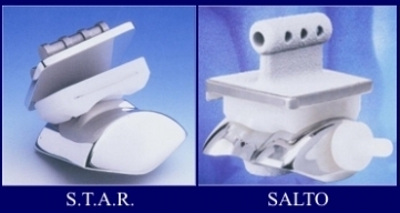
We currently use three different prosthesis models: on the one hand the S.T.A.R. prosthesis from the company SBI, on the other hand the SALTO prosthesis or SALTO-Talaris prosthesis from the company Tornier.

More than 80% of the patients operated on by us are satisfied or very satisfied with their operated ankle joint and would have this operation repeated at any time.
However, about 10% of patients continue to have complaints of various kinds and are unsatisfied with the surgical result.
The range of motion in the upper ankle joint was improved by an average of 8°, with a clear correlation to the preoperative range of motion. Complications can occur during and immediately after any surgery. For example, malleolus fractures (ankle fractures) or wound healing disorders, which must then be treated accordingly. In our patient population, this complication rate was 7%. In some patients it was necessary to perform so-called revision surgery for various reasons. Causes may include loosening of individual prosthesis components, formation of periprosthetic cysts, malpositions, fractures, and persistent pain. Such follow-up operations (approx. 12% in total) include, for example, the replacement of individual prosthesis components, the filling of cysts with autologous or foreign cancellous bone, or position corrections in the ankle joint. In a total of 60 patients, however, the implanted endoprosthesis had to be removed and subsequently a fusion operation performed. Due to the very sparing bone resection during prosthesis implantation, subsequent fusion is possible as a so-called retraction operation.
Course of Treatment
The implantation of an OSG-TEP is performed under regional or general anesthesia. This generally lasts between 60 and 90 minutes. If there are no complications, the patient can leave the bed one day after the operation with the help of physiotherapy. The ankle joint is exercised with physiotherapy from the 1st day, and manual lymphatic drainage is also performed to reduce swelling of the joint.
After the wound has healed (10 to 12 days), the stitches are removed and a circular cast (plaster) or orthosis is applied.
The patient can now leave the clinic and put full weight on the leg with the orthosis in place. After about 6 weeks, the prosthesis has largely grown together with the bone. After a clinical and radiological examination in our outpatient clinic, intensive physiotherapeutic treatment is carried out. This is preferably carried out on an inpatient basis as a follow-up treatment, but can also be carried out on an outpatient basis at the patient's home.
The rehabilitation phase takes about 12 weeks postoperatively.
Full weight-bearing capacity of the operated ankle should be achieved within the first 6 to 12 months. Normally, after completion of the treatment, the patient can walk without complaints and resume everyday tasks and activities.
You should consult your attending physician about resuming professional activities. Extreme loads, e.g. jumping from great heights, martial arts, etc. should be avoided to protect your joint. There is nothing wrong with moderate sporting activity.
There are no major studies available on the long-term prognosis and durability of prostheses. In the meantime, some authors have given so-called service lives of 80-90% after 5 years or 70-80% after 10 years. Larger literature studies (meta-analyses) on the results of ankle prostheses have been published, for example, by Stengel et al.(2005), Haddad et al. (2007), Gougoulias et al. (2010) and Zhou et al. (2011).
The results from various studies from centers in Europe and the USA with a total of 5000 ankle joint endoprostheses were evaluated here.


